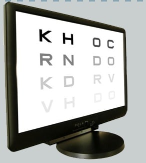
For many years contrast sensitivity testing has been recognized to be an useful strategy to assess visual function in many ophthalmological diseases as glaucoma, optic neuropathies, amblyopia as well as in neurological and, recently, even psychiatric conditions.
So far as we know, available contrast sensitivity tests are performed by means of paper-made screens or backlighted windows and do not employ a psychophysical procedure to assess the exact threshold value for every tested spatial frequency.
 The new VistaVision Contrast Sensibility Test© (DMD Med Tech, Turin, Italy) is a computer-based psychophysical tool, using a particular staircase procedure to fit the psychometric function.
The exam evaluates spatial frequency ranging from 0.75 c/deg to 18 c/deg, widely covering the values processed by the magnocellular (under 1.3-2 c/deg, according to Legge,1978) and parvocellular ganglionar pathway.
Within this range, the VistaVision evaluates contrast sensitivity across six different spatial frequency values (0.75, 1.5, 3, 6, 12, 18 c/deg). Test distance is 3 meters. Subjects are presented sinewave gratings (mean luminance 120 cd/m2) in random order, each oriented along the vertical or tilted left/right side and are asked to report the orientation of the displayed grating by a remote control. For each spatial frequency value, threshold level is obtained by a 4-2 staircase procedure.
The difference between VV function and a 3-forced choice paradigm (3AFC) relies on the further possibility to answer “null” by clicking an appropriate button. X normal subjects aged x year (±y) were recruited. Inclusion criteria were BVCA: 60/60, ametropy <±3 diopters, absence of ophthalmological and systemic diseases.
Exclusion criteria were intraocular pressure higher than 17 mm Hg (G) or inheritance for glaucoma, strabismus, amblyopia, diabetes, systemic therapies able to induce visual field defects, cognitive disability and bad collaboration. The exam was performed monocularly.
Every patient showed foveal fixation as tested by Cuppers’ visuscope.
An high correspondence both in the shape and in the relative threshold values is found between the obtained spatial contrast sensitivity function and the reference one. To account for any age effect, a correlation analysis has been performed by Pearson test. A negative significant correlation is found between age and contrast sensitivity above 12 c/deg.
The question arises if this correlation may be accounted simply for the mild age-dependent reduction of the media transparencies. To investigate this point, the correlation of the two higher spatial frequencies tested has been compared. Indeed, since ocular media light absorption is proportional to the age and mainly the higher frequencies are affected, then an higher correlation with the 18 c/deg as compared to the 12 c/deg is expected to occur.
Actually, it was not the case and the age-spatial frequency correlation resulted to be stronger for 12 c/deg than for 18 c/deg. This suggests that media changes due to ageing account for the CS decrease mainly at high spatial frequencies whereas other factors, namely the retinal tissue involution, would affect contrast sensitivity at lower spatial frequencies. In any case an ageing effect seems to worsen contrast sensitivity for spatial frequencies above 6c/deg.
In conclusion, VistaVision contrast sensitivity test is a fast, precise and reliable device to assess this function in ophthalmological and neurophthalmological field. Mean examination duration resulted to be less than 2 minutes per eye, and test-retest variability was negligible. The obtained psychometric curve fits well in shape and values with data reported in literature and has the advantage to examine a wider range of frequencies compared to the other current instruments. Consistently with previous studies, contrast sensitivity decreases as a function of age at the higher tested frequencies.
Carlo Aleci, MD, PhD
Ophthalmology Department, The Gradenigo Hospital, Turin, Italy
carlo.aleci@gradenigo.it
